Bernard J Gersh, MB, ChB, DPhil, FRCP, MACC
- Editor-in-Chief — Cardiovascular Medicine
- Section Editor — Coronary Heart Disease; Myopericardial Disease
- Professor of Medicine
- Mayo Clinic College of Medicine
- It may occur in one or both legs depending on the location of the clogged or narrowed artery. Other symptoms of PVD may include: Changes in the skin, including decreased skin temperature, or thin, brittle, shiny skin on the legs and feet. Weak pulses in the.
- Thus, examination of the carotid pulse provides the most accurate representation of changes in the central aortic pulse. The brachial arterial pulse is examined to assess the volume and consistency of the peripheral vessels. UNEQUAL OR DELAYED PULSES. Inequality in the amplitude of the peripheral pulses.
Peripheral pulses were graded as either absent or present. Absent was graded as 0⁄3, present but reduced (1⁄3), normal (2⁄3) or bounding (3⁄3). Femoral bruits were graded as.
Catherine M Otto, MDCatherine M Otto, MD
- Editor-in-Chief — Cardiovascular Medicine
- Section Editor — Cardiac Evaluation; Valvular Disease
- Professor of Medicine
- University of Washington
Susan B Yeon, MD, JD, FACC
- Deputy Editor — Cardiovascular Medicine
INTRODUCTION
Assessment of the arterial pulse characteristics is an integral part of the cardiovascular examination. The arterial pulse examination and abnormalities caused by cardiovascular disease are discussed in this topic.

Examination and evaluation of lower extremity and upper extremity peripheral arterial disease are discussed separately. (See 'Clinical features and diagnosis of lower extremity peripheral artery disease', section on 'Pulses' and 'Overview of upper extremity peripheral artery disease', section on 'Presentation'.)
EXAMINATION COMPONENTS
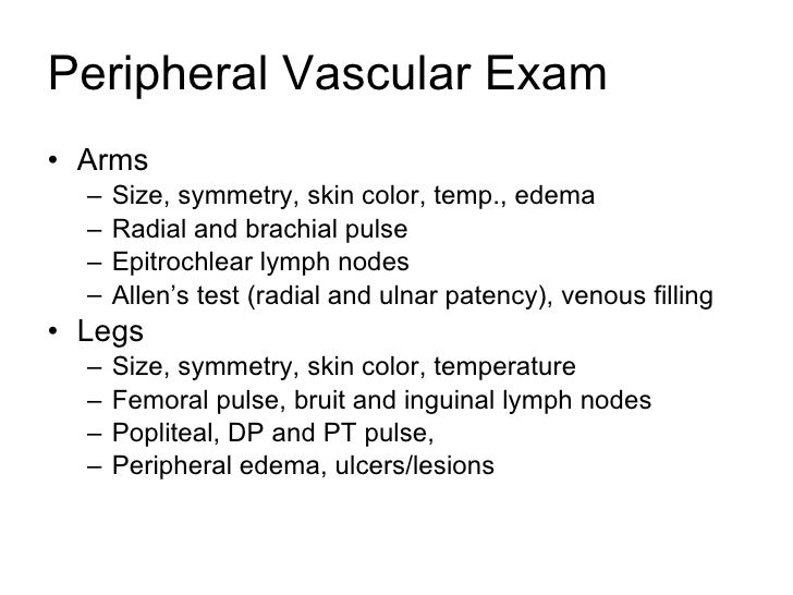
Carotid, radial, brachial, femoral, posterior tibial, and dorsalis pedis pulses should be routinely examined bilaterally to ascertain any differences in the pulse amplitude, contour, or upstroke. Popliteal pulses should also be examined when lower extremity arterial disease is suspected. Adobe flash for firefox mac os.
NORMAL EXAMINATION
The carotid pulse contour is very similar to that of the central aortic pulse; a delay in the onset of the ascending limb of the carotid pulse, compared with the central aortic pulse, is only about 20 msec. Thus, examination of the carotid pulse provides the most accurate representation of changes in the central aortic pulse. The brachial arterial pulse is examined to assess the volume and consistency of the peripheral vessels.
UNEQUAL OR DELAYED PULSES
Inequality in the amplitude of the peripheral pulses may result from:
Subscribers log in here
Part II: Assessment Techniques
Inspection
As you prepare to begin the actual assessment, you already have obtained and recorded the patient history and you arm yourself with pertinent data such as their chief complaint and allergic history.
Also keep in mind to allow a certain amount of time in order to complete a thorough exam. Many nurses do not have large blocks of time for completion of the assessment but you must be as thorough as possible. If this is an admission assessment, you must allow enough time to be complete. If this is an on-going assessment, not as much time will be required.
Begin Exam
- Patient undresses, but allow for privacy.
- Have the patient sit upright and inspect the thorax from the front.
- Now inspect from the back of the patient.
You will inspect for posture and symmetry of the thorax, color of the skin, gross deformities of the skin or bone structure, the neck, face, eyes, and any abnormal contours. Breathing patters will also be noted. Be especially aware of the presence of cyanosis. Central cyanosis is a condition which will cause the lips, mouth, and conjunctiva to become blue. Peripheral cyanosis will cause blue discoloration mainly on the lips, ear lobes, and nail beds. Peripheral cyanosis might indicate a peripheral problem of vasoconstriction, and would generally be less severe than central cyanosis, which could indicate heart disease and poor oxygenation.
Thorax Logo soft for mac.
Inspect for symmetry of thorax, point of maximum intensity (PMI). PMI is easier to find if the patient will lay on the left side. PMI may also be palpated. Check skin color of thorax.
Eyes
Arcus Senilis is a light gray ring surrounding the iris, common in older patients; in younger patients it might indicate a type of lipid metabolism disorder, which is a precursor to coronary artery disease.
Xanthelasma is yellowish raised plaques on the skin surrounding the eyes. Can also appear on the elbows. This is a possible indication, or sign of hypercholesterolemia, often a precursor to coronary artery disease (atherosclerosis).
Palpation
Palpation, or touching, is the next part of the exam. In the stop above, if we noted any abnormalities, we will now palpate and evaluate them further.
Skin: temperature, texture, moisture, lumps, bumps, tenderness.
Examination of extremities for edema might also indicate a cardiovascular problem. Examine the feet, ankles, sacrum, abdomen, trunk, and face for edema. If you notice puffiness of frank edema, then palpate the area for pitting edema. Most facilities recognize the following scale:
+1 Pitting Edema | = | 0 to ¼ inch indentation |
+2 Pitting Edema | = | ¼ to ½ inch indentation |
+3 Pitting Edema | = | ½ to 1 inch indentation |
+4 Pitting Edema | = | More than 1 inch indentation |
Breathing: lay hands the chest at different locations and feel the respiratory patterns, feel the ribs elevate and separate during normal breathing.
Peripheral Pulses +3
Pre-Cordial Areas you can feel the pounding of the heartbeat, normal and abnormal pulsations o the chest wall; PMI, as mentioned above.
Arteries: Assess all pulses
You undoubtedly assessed the apical pulse earlier when you took the patient’s vital signs, if not, now is the time. Assess the following pulses:
- Apical heart rate – monitor for a full minute, note rhythm, rate, regularity.
- Radial pulse – monitor for a full minute. Note the rhythm, rate, and the regularity. Note any differences from right to left radial, a large difference might indicate arterial blockage or even enlarged ventricles. If pulse is regular but volume diminishes from beat to beat, this might indicate left-sided heart failure and is called pulses alternans. If the volume of the pulse diminishes on inspiration, might indicate constrictive pericardial disease, the condition is called pulsus paradoxus.
- Carotid, brachial, femoral, popliteal, posterior tibialis, and dorsalis pedis pulses – when checking these pulses do it the same way as the others mentioned in this section; right then left side. When you check the carotid, press gently and do not rub.
Do not palpate carotid on persons with known carotid disease or bruits; listen with stethoscope instead; and do not palpate both carotid pulses at the same time.
Carotid Artery:
- Plateau pulse – slow rise and slow collapse pulse; may be caused by aortic stenosis, slow ejection of blood through a narrowed aortic valve.
- Decreases amplitude (grade point pulse) – due to hemorrhagic shock, pulse is weak due to decreased blood volume.
Bounding Pulse - (Grade IV) can be due to hypertension, thyrotoxicosis, others; associated with high pulse pressure, the upstroke and downstroke of the pulse waves are very sharp.
It is common to use +1, +2, etc. when recording pulses:
Peripheral Pulses 3+
- 0 = absent
- +1 = diminished or decreased
- +2 = normal pulses
- +3 = full pulse or slight increase in pulse volume
- +4 = bounding pulse or increased volume
Peripheral Pulses Grading
Veins – neck, arms, legs, etc.
Next: Part II: Assessment Techniques, Con't.
